Children exposed to adult radiation levels from CT scans
Research shows that the radiation doses given to children in CT scans in the1990s were significantly higher than today.
CT SCANS: Radiation doses need to be adjusted downwards when small bodies get a CT scan. We have little evidence of what the CT scan radiation doses were for children in the 1990s.
“Due to the technical limitations of CT scanners in the 1990s, it was common to use the same device settings for children and adults,” says assistant professor Benthe Toft at NTNU’s radiography programme in Gjøvik.
Results from Toft’s Master’s thesis suggest that young children may have been given two to three times higher organ doses than adults. Now researchers with the International Pediatric CT scan study (EPI-CT) are investigating whether a possible link exists between radiation doses from childhood CT scans and later cancer.
Children are more sensitive
Despite clear benefits as a medical tool, CT scans pose a potential risk. Scientists assume that children are more sensitive to radiation than adults, but are not certain how much radiation people can tolerate before it causes late effects.
Doses are determined by a person’s weight and size, and when children need to have a CT scan, the dose is reduced. At the same time, a certain minimum dose of radiation is needed to obtain quality images.
“Toft’s findings reflect the technological developments that have made it possible to obtain good images with less radiation. The dose that was used earlier was probably necessary in order to create images that could be interpreted and used in the clinic,” says Erik Magnus Berntsen, associate professor at NTNU’s Department of Circulation and Medical Imaging.
- You might also like: Mirror, mirror, will I have a heart attack?
Scanty information
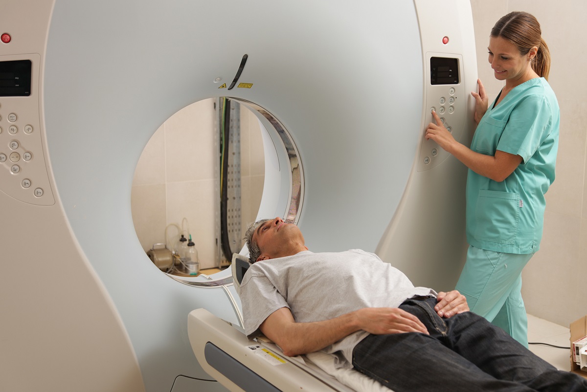
Adults and children were often given the same radiation doses. Illustration photo: Colorbox
Prior to technology’s digital advance, radiation protocols were conducted manually.
“As hospitals replaced their CT scanners, they also updated their protocols, and that means we generally have only limited historical protocol records for children who were scanned,” explains Toft.
The Norwegian Radiation Protection Authority (NRPA) did, however, conduct a national survey in the 1990s. Based on this information, Toft has tried to recalculate the radiation dosages that children received. She has based her calculations on new software for calculating organ doses in children and adolescents, which was developed for the multinational EPI-CT epidemiological research project. Her work is an important contribution to the development of the project’s dosimetry, and with the project partners, has been published in the journal European Radiology.
Awaiting results
Toft estimates that the youngest children may have received significantly higher doses than what is common today. The consequences of this are not yet known, and this is what EPI-CT is investigating.
By reconstructing earlier doses with the dosage calculator Toft used, the researchers will examine a possible correlation between childhood CT examinations and later cancer. The first results are expected in 2017.
“Based on our results, the CT scan doses given to children in the 1990s were of a magnitude that we can expect results in the EPI-CT project,” says Toft.
Need for standardized doses
Berntsen says that Toft’s main finding, that the organ dose at that time was two to three times greater than today, seems plausible. “This is mainly due to the technological developments that have resulted in improved CT scanners with smaller radiation doses. The findings are important and useful for epidemiological studies of increased cancer incidence as a result of earlier CT scans,” he said.
The EPI-CT project will help optimize protocols and standardize doses nationwide, enabling CT scan procedures to be adapted to children.
“If we decrease the dose too much, we lose image quality. The radiographer’s job is to provide good visual material that enables a diagnosis of the medical issue, at the same time making sure that the radiation dose for children isn’t too high,” says Toft.
Sources:
• Olerud HM, Toft B, Flatabø S, Jahnen A, Lee C, Thierry-Chef I (2016) Reconstruction of pediatric organ doses from axial CT scans performed in the 1990s – range of doses as input to uncertainty estimates.European Radiology.Vol. 26, Issue 9, pp 3026–3033
• Mathews John D, Forsythe Anna V, Brady Zoe, Butler Martin W, Goergen Stacy K, Byrnes Graham B et al. Cancer risk in 680 000 people exposed to computed tomography scans in childhood or adolescence: data linkage study of 11 million Australians BMJ 2013; 346 :f2360
• Pierce MS, Salotti JA, Little MP, McHugh K, Lee C, Kim KP et al. (2012) Radiation exposure from CT scans in childhood and subsequent risk of leukaemia and brain tumours: a retrospective cohort study. Lancet 380(9840):499–505