Norwegian ultrasound technology in Cape Town
Norwegian researchers have installed a system that uses 3D ultrasound and image guidance in one of Africa’s biggest children’s hospitals. This could make it easier to treat brain diseases in children.
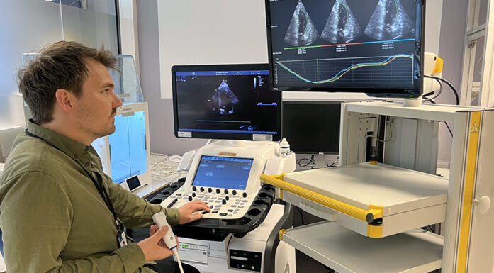
SINTEF researchers Tormod Selbekk and Reidar Brekken both work with ultrasound on a daily basis and their doctoral degrees are in ultrasound and its clinical applications.
They are currently working as part of a specialist team in Trondheim, which is one of the most advanced locations in the world in its use of ultrasound and 3D image guidance during brain surgery. Researchers, engineers and doctors at St Olav’s Hospital have been working together for many years, and have produced some excellent results.
FACTS:
- Researchers at SINTEF and the Norwegian University of Science and Technology (NTNU) have been working with doctors at St Olav's Hospital for many years, via the National Centre of Expertise for ultrasound and image-guided treatment.
- Now the Trondheim specialists are working with the Division of Neurosurgery at the Groote Schuur Hospital at the University of Cape Town, and at the Red Cross War Memorial Children's Hospital.
Start of the partnership
But how did their technology get to Cape Town?
It started eight years ago with a SINTEF workshop in the city – a gathering of medical and ICT specialists, including staff from the Division of Neurosurgery in Cape Town, whom Tormod Selbekk then met for the first time. One of them was Graham Fieggen, a professor at the University of Cape Town, and head of the Division of Neurosurgery at the acclaimed Groote Schuur hospital. He also runs the Department of Neurosurgery at the Red Cross War Memorial Children’s Hospital in the city.
“We had some interesting conversations, but didn’t agree on working together then and there”, relates Selbekk.
However, one day in January 2013, an e-mail arrived from South Africa.
“Fieggen had checked our publication list, and saw that we were still heavily involved in research into ultrasound and brain surgery. He also knew a paediatric neurosurgeon who wanted to take a doctoral degree in the use of ultrasound on children”, smiles Selbekk, who is now supervisor for that young doctor.
Llewellyn Padayachy travelled to Trondheim twice in 2013 to look into what the Norwegian researchers were studying. He also had his own ideas about how ultrasound imaging could be used to measure pressure in the brain.

The Norwefgian scientists Tormod Selbekk (t.v.) og Reidar Brekken together with Llewellyn Padayachy who is pediatric nevro surgent by Red Cross War Memorial Children’s Hospital.
Many children with brain injuries
Traffic accidents are extremely common in South Africa and the Red Cross Hospital admits children from most of the country. It is also a treatment centre for children with traumas, those who have suffered blows to the head, and those who have diseases that cause fluid to build up on the brain.
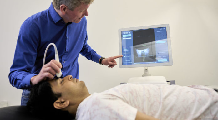
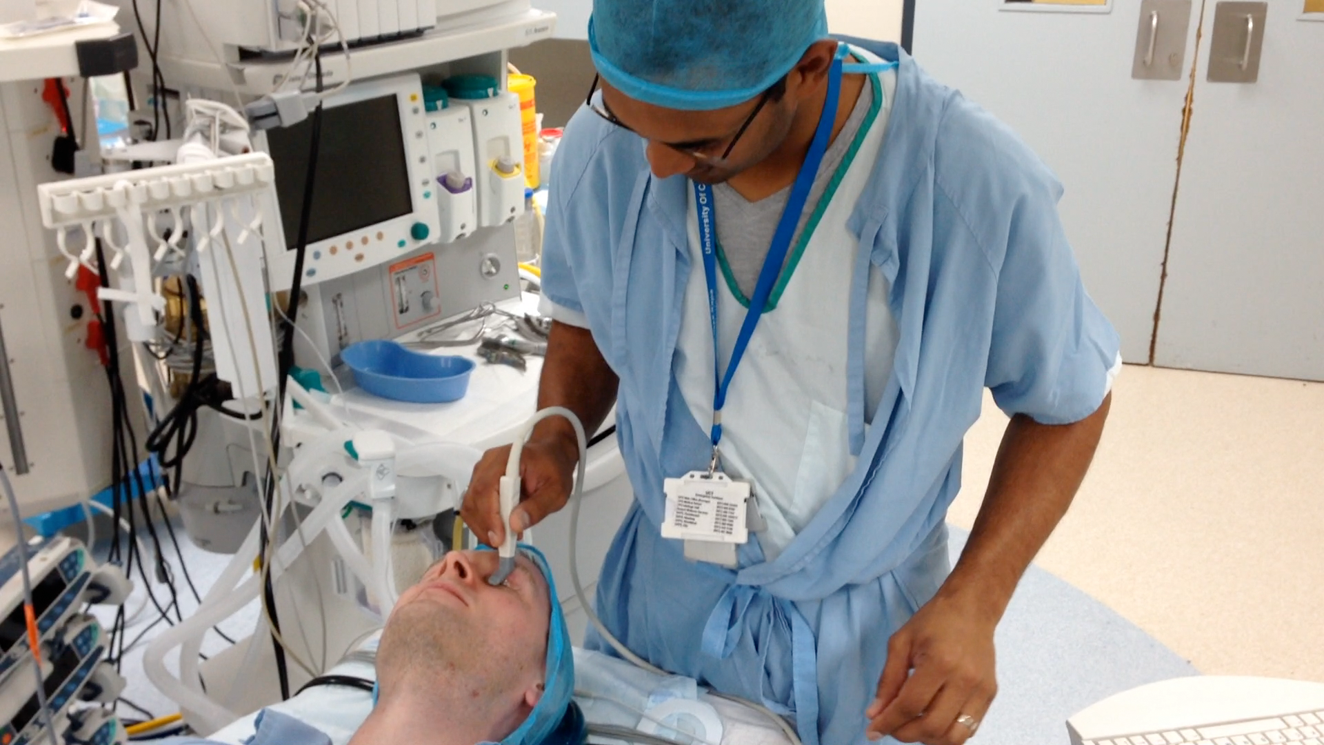
In all these cases, it is important to take measurements of the brain in order to identify injuries. The use of ultrasound means that it isn’t necessary to drill holes in the cranium in order measure whether there is high pressure on the brain.
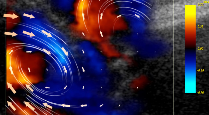
By processing the ultrasound images, the Norwegian researchers have helped to develop methods that can quantify movements in brain tissues. They have also installed a system that makes it possible to generate 3D images of the brain using ultrasound. This allows them to determine whether it will be possible to use this approach to provide better diagnosis and treatment of medical conditions in children.
“We went down there in April to install the system which allows them to collect 3D images. Installing the system also means that it will be possible for us to carry out more work with the specialists in Cape Town”, says Reider Brekken.
This is the system the researchers have installed:
- The CustusX software has been developed by SINTEF, NTNU and the hospital, assisted by funds from the National Centre of Expertise for ultrasound and image-guided treatment. This software makes it possible to put together ultrasound images in volumes that allow us to navigate in 3D. The programme has been used in the Netherlands, but this is the first time the tool has been permanently installed in another country.
- The system set up at the Children's hospital in Cape Town consists of a Mac computer installed with the CustusX software. This is connected to both an ultrasound scanner and a tracking system that acts as a GPS and can track the tiny sensors that are fitted to the ultrasound probe. The system can thus keep constant track of the probe that is sending and receiving the sound waves.
- Using this system, the surgeon obtains 2D images of the brain, which he can convert into 3D images ready for analysis.
No need to drill holes
Two partnership projects have now become a reality. The first involves the ultrasound imaging of the optic nerve in children, in order to measure pressure on the brain. By holding the probe against a child’s eye socket, doctors are able to obtain ultrasound images and information. This can simplify a diagnosis, and avoid the need to drill holes and insert pressure gauges.
The second project is looking into whether it is possible to use ultrasound to guide the insertion of small drains (tubes) into cavities in the brain in order to drain fluids, in diseases such as hydrocephalus.
Images from the children’s hospital in Cape Town are now sent to Trondheim, where they are used as material for future research.
“This is an exciting partnership”, says Reidar Brekken. “To do clinical research, you need a lot of patients. Since Red Cross is one of the biggest children’s hospitals in Africa, this is something they can provide. We give them technological assistance and they give us material for further research – including how to obtain more information from the ultrasound images than we are currently able to.